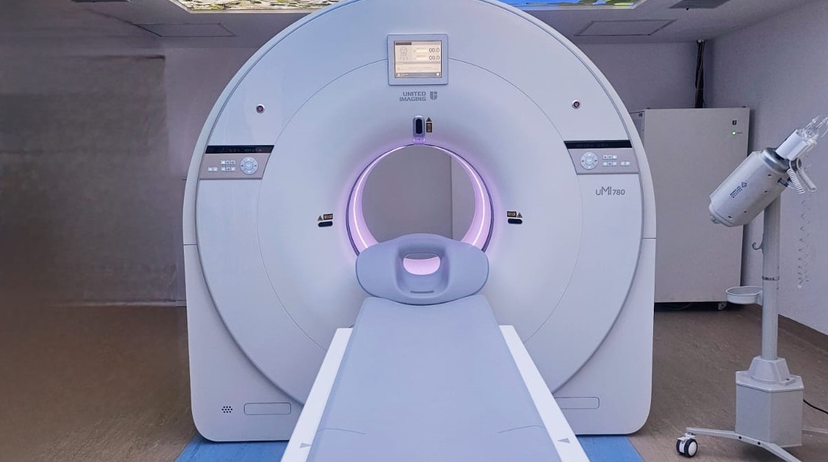
Digital PET Scans: The Game Changer in Cancer Screening
What is PET Scan?
Positron emission tomography (PET) is a nuclear medicine technique that gauges the metabolic activity of cells in body tissues. Typically employed by oncologists, neurologists, neurosurgeons, and cardiologists, PET’s applications are expanding due to technological advancements. It may complement other diagnostic tests like computerized tomography (CT) or magnetic resonance imaging (MRI), and the integration of PET and CT or MRI in a single scanner, known as PET-CT or PET-MRI, holds significant potential for diagnosing and treating cancers.
Why is PET performed?
In general, PET scans may be used to evaluate organs and/or tissues for the presence of disease or other conditions. PET may also be used to evaluate the function of organs, such as the heart or brain. The most common use of PET is in the detection of cancer and the evaluation of cancer treatment.
More specific reasons for PET scans include, but are not limited to, the following:
- Detecting cancer.
- Revealing whether your cancer has spread.
- Checking whether a cancer treatment is working.
- Finding a cancer recurrence.
The role of a PET scan in cardiology
The role of a PET scan in cardiology is significant in assessing various aspects of heart health. A heart PET scan is capable of determining whether specific regions of the heart muscle are adequately supplied with blood, identifying the presence of heart damage or scar tissue, and detecting the accumulation of abnormal substances in the heart muscle.
Cardiac PET scans are typically performed when medical professionals need to ascertain specific information about the patient’s heart condition. These situations include determining which areas of the heart are experiencing insufficient blood flow, diagnosing coronary artery disease, assessing the condition of the heart muscle, detecting potential heart infections, identifying cardiac sarcoidosis, evaluating the suitability of a procedure, measuring the extent of heart damage following a heart attack, assessing the effectiveness of a cardiac treatment plan, and gauging the overall health of the heart before a medical procedure.
How does a PET scan work?
A PET scan uses a radiotracer in the form of radioactive sugar. The body’s cells take in different amounts of the sugar, depending on how fast they are growing. Cancer cells grow quickly, meaning they are more likely to take up larger amounts of the radiotracer.
A PET scan can identify regions where the radiotracer accumulates or doesn’t, providing insight into the functioning of specific cells. At healthcare facilities such as The Nairobi West Hospital, a PET scan technologist administers the radiotracer intravenously through a vein
The radiotracer takes some time to make its way through a person’s body. The individual then slides through the central hole in the PET machine before the scan takes place.
When the radiotracers in a PET scan decay, they produce small particles called positrons, which react with electrons in the body. When they combine, they annihilate one another, producing a small amount of energy in the form of two photons. The detectors in the PET scanner measure these photons, which they use to create images of inside a person’s body. Cancer cells show up as bright spots on PET scans because they have a higher metabolic rate than do typical cells.
How does a person prepare for a PET scan?
When preparing for a PET scan, a person may have to stop eating or drinking for 6 hours before the procedure.
An individual should also inform the doctor or nurse about any medications they are taking, including any allergies the person has and whether they have had previous reactions with nuclear medicine scans.
What happens during a PET scan?
At The Nairobi West Hospital, like in many healthcare facilities, PET scans often take place in the radiology or nuclear medicine department. A typical PET scan involves the following steps:
A person will need to remove all jewelry or metal items that could interfere with the scan. They may be able to wear their own clothes, though sometimes they may need to wear a gown during the test.
A doctor will begin by administering the radiotracer. They will most often do this intravenously using a needle.
The PET scanner has a hole in the middle. A person will lie on a padded table that moves through the hole of the scanner.
A technician may ask the person to change positions to allow them to take scans of different views. Other than during those movements, an individual needs to remain still during the scan.
What can a person expect after a PET scan?
PET scans are usually carried out on an outpatient basis. This means you won’t need to stay in the hospital overnight. You should not experience any side effects after having the scan.
The Nairobi West Hospital usually issues results within 24-48 hours and discusses with the patient at their next appointment.
What are the possible risks of a PET scan?
A PET scan involves a slight risk of radiation exposure due to its use of radioactive materials. However, the overall radiation dose is comparable to that of a routine chest X-ray or CT scan. Although there are concerns about potential cancer risks from low radiation levels, the risk associated with PET scans is relatively small, and the diagnostic benefits typically outweigh it.
Medical professionals in nuclear medicine strive to minimize radiation exposure by using the least amount of radiotracer necessary for effective imaging. The radiotracer’s radioactivity diminishes over time, and it usually exits the body naturally within a few hours. Drinking plenty of fluid after the scan can help flush it from your body.
As a precaution after a PET scan, it is advisable to avoid close contact with pregnant women, babies, and young children for a few hours, as the individual may emit slight radiation during this period.
For PET Scan services, The Nairobi West Hospital provides assistance; contact them at 0730 600000 or visit their location on Gandhi Avenue in Nairobi.

